

The Image Companion® Short Overview:
Menus:
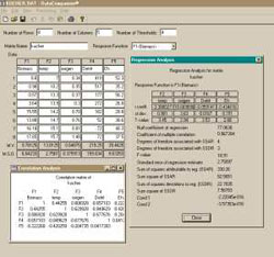
Screenshot of Data Companion® Menu and selected Processing results
Functionality:
Preprocessing
- LUT/Enhancing/ROI
- Automatic and Manual Stretch
- Filtering Functions
- Edge Detection
- Edge Enhancement
- Sharpening Functions
- Dilation Functions
- Manual Editing
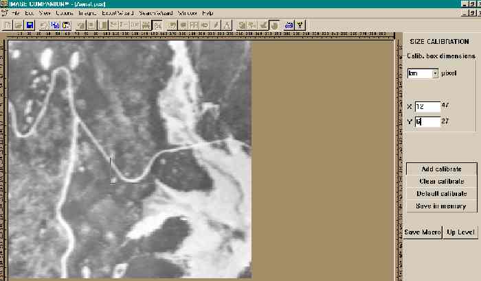
- Calibrating
- Special Functions (Activate Object, Delete Activated Object)
- Image Annotation
- Image Draw
Processing
- Manual Segmentation
- User Guided Semi-Automatic Segmentation
- Single Layer Segmentation
- Multi Layer Segmentation
- Measuring
- Sorting
- Profiling
- Histograms
- Statistics
- Search for Object(s)
- Similarity Indexing
Postprocessing
- Search for Template
- Search for Multi-Component Template
- Export to Excel
- Generic Export
- Macro Library
- Batch Processing
- User Defined Measuring
- Image Format Conversion
System Highlights:
System Requirements
- Intel PII or higher
- 64MB RAM minimum
- At least 800x600 pixels resolution with 256 or higher colors support
- Windows XP, Windows NT/2000, Windows 7/8/10
Companion Product
- Data Companion® provides for full correlation, regression and multifactor analysis
- Unique statistical weights analysis
- Can exchange data with Image Companion® through a Metafile
Special Promotions
- Government, Volume, Educational and Academic discounts
- Installment Financing with verified credit
- Discount on customization
- 30-days money back
- Quotation through the Web site.
Sample Results:
Samples of Image Companion® screenshots with:
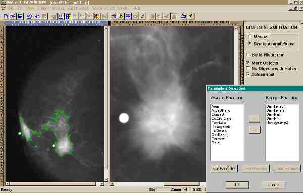
Mammography: Computer-Aided Detection in Radiology: Mammographic image automatically processed with the user defined objects settings (through multi-thresholding) and suspicious areas are clearly marked and quantified. Such areas can be compared with enhanced view of the tumor area (right part of the display window).
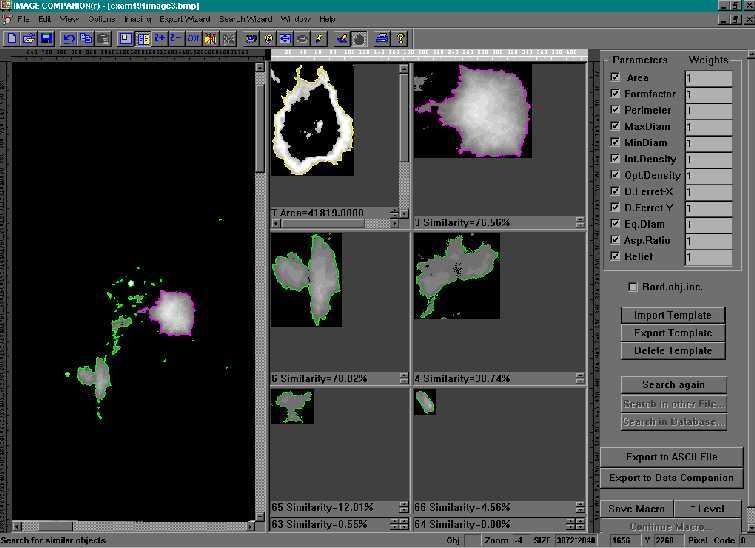
Mammography: Indexing and retrieval from database: Previously detected suspicious areas are compared with cancer template imported from the other cases. All areas of the processed image ranked based on calculated similarity to the template (selected by the user).
Cytology: Lab Automation in Microscopy: Blood count and full statistics are performed using automatic image processing in batch using Macro Library previously saved by the user.
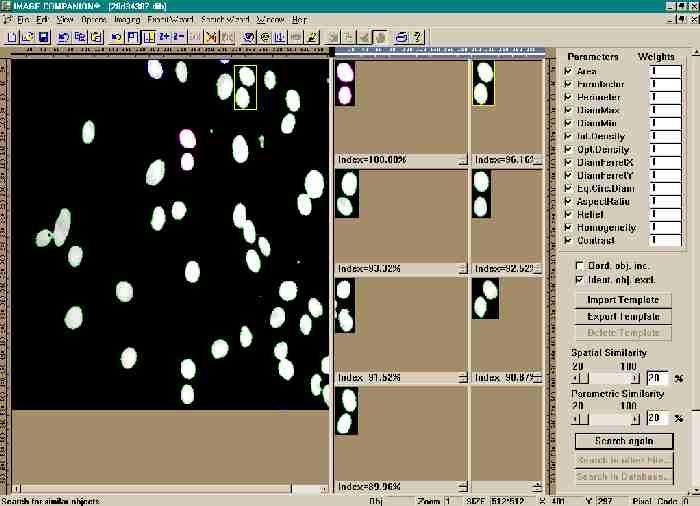
Cytology: Search within processed file: Previously detected objects are compared with selected by the user template object. All areas of the processed image ranked based on calculated similarity to the template. Numeric results are prepared for exporting, new template is saved for future use.
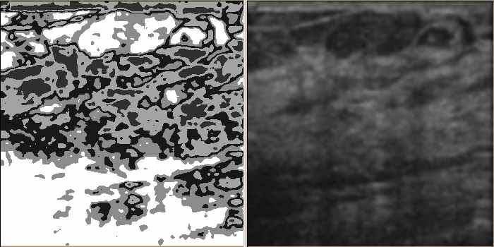
Ultrasound: Computer-Aided Detection in Ultrasound: The current method of detection and analysis of suspicious areas is based on manual inspection of the acquired images. It is subjective and error-prone. Our technology not only can view and display comprehensively (enhanced view on the left and traditional view on the right), but can detect, quantify and search images too.
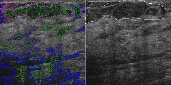
Ultrasound: Quantification and Scoring of suspicious area: Previously detected suspicious areas are automatically detected and quantified. They can then be compared to one marked by radiologist (accuracy test) and numerically scored to the level of suspicion.